3 TESLA MRI
3 TESLA MRI- A CLINICAL ADVANTAGE
An MRI examination utilizes several kinds of magnets, pulses of radio frequency (RF), and sophisticated and powerful computers. Images are generated based on the physics involving the excitation and relaxation of millions of spinning protons found in water molecules inside the human body.
The image taken with the MRI begins and ends with the computer. It directs all the activity of getting the image, sending it to gradient amplifiers and RF transmitters, turning them on and off to obtain the proper pulse sequence to the Magnet, passing the data through the RF amplifier to a data converter that digitizes the signal and back to the computer to be reconstructed into an image.
The first MRI was performed on a human being in 1977 and at that time it took almost five hours to produce the image. Thank god the scenario is completely different now. With the advent of newer technologies we are able to produce images faster, with better details and using less injectable contrast
The most common MRi macine in clinical use is 1.5 T (tesla) unit. The T refers to tesla which is a measure of the field strength of the machine. Therefore, if 1.5 T is the most commonly used and 3T is the newest approved, is the new approved 3T machine actually improved? YES would be a reasonable answer. The 3T is faster, meaning greater patient tolerance. As a result, there are fewer artifacts because of motion. It is particularly important when studying children, where a request to remain motionless may go unheeded unless they receive medication. So, another advantage may be less use of sedation. The 3T MRI produces a higher spatial resolution and image contrast without injection of contrast medium, resulting in better visualization of anatomic structures. Thus, if a picture is worth a thousand words, the 3 Tesla MRI is an encyclopedia. Subtle abnormalities can more easily be detected. The image can be so good that the radiologist may not require intravenous gadolinium or intra-articular contrast medium administration.
So, what is it about the 3T MRI that makes it worth having? While there are financial considerations for a radiology facility, it is acceptable to presume that for neurological, musculoskeletal and cardiac images, your best choice is the 3T. It may reduce imaging time and/or improve resolution and contrast and give thinner slices. Some of the most advanced imaging techniques are available with 3T MRI. In the era of advanced management tools available(Robotic surgery) it becomes mandatory to have such high end MRI machines to get quality imaging. This aids in providing utmost precise imaging findings and diagnosis which would help in diagnosis and management.
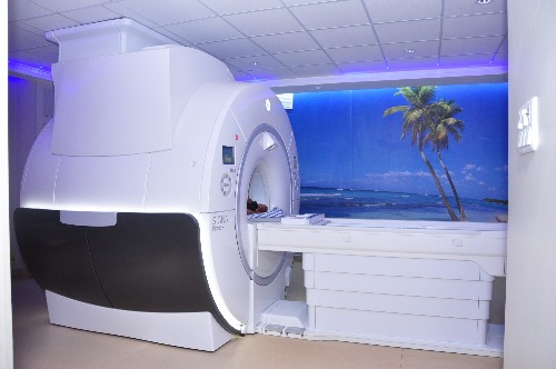
With the acquisition of the 3 Tesla MRI, the Yenepoya Medical College Hospital becomes the fourth Teaching College in the country to have this advanced facility. This is going to be highly beneficial not only to the hundreds of patients who visit the hospital for treatment but also expose the students to the most advanced technologies available today.